If you run a lab that still reads at the microscope, digital pathology probably reaches you as a conversation about scanners: brands, speeds, load capacity. That conversation matters, but it isn't the one that decides whether going digital works.
What decides it is something else: whether each scanned slide finds its case on its own, where hundreds of gigabytes a month get stored, whether the site's internet link can keep up with uploading them, and what changes in the pathologist's day. This guide walks through those four questions with measured figures, not promises.
In short
- A digital slide (whole slide imaging) is the complete image of a slide at several resolution levels, which the viewer loads piece by piece depending on where you look.
- In Histeva's technical tests, slides scanned at 20x came in at 0.2 to 0.5 GB each in DICOM.
- Histeva stores slides in DICOM WSI, an open standard, and converts the SVS, NDPI, MRXS, iSyntax and CZI formats at the site.
- The scanned slide links to its case automatically through the DataMatrix code on its label. If it can't find the case, it goes to a reconciliation queue.
- You can start with the microscope and scan later: the codes are the same, so there is no migration.
- Image-analysis AI is a future phase of Histeva. Today the AI works on text.
What a digital slide is and how large it is
A slide scanner photographs the entire slide at high resolution and saves the result as a pyramid: the full-resolution image at the bottom, and progressively smaller versions on top. Each level is split into tiles, small squares the viewer requests only when they appear on screen. That's why you can open a slide of hundreds of megabytes and navigate it like a map, without downloading the whole thing.
File size depends on the scanner, the format and the magnification. In Histeva's tests with public slides scanned at 20x, an Aperio or Hamamatsu slide came to about 0.2 GB in DICOM, and a 3DHistech slide to 0.53 GB. Slides at 40x are larger, and Histeva has not yet published a measured figure for that magnification. If someone quotes 2 GB per slide as a general rule, ask them to measure it with your slides.
The cost of storing a slide turned out to be small. The bottleneck is elsewhere: the lab's upload link.
Bandwidth, the math nobody does
A lab that scans 200 slides a day, at 0.2 to 0.5 GB per slide, generates between 40 and 100 GB a day. Uploading that within 24 hours takes between 3.7 and 9.3 Mbps sustained around the clock, and twice that if you only upload during business hours. With upload links of 4.5 to 8 Mbps, like the ones measured in the tests, a 200 MB slide takes between 3 and 6 minutes to upload.
That's why the Histeva Edge uploads in the background, with a queue that puts intraoperative cases ahead of urgent ones and urgent ones ahead of routine work. If an upload is interrupted, it resumes from the last confirmed part, and it keeps the original file at the site until the cloud confirms it was received. You can also set time windows for uploading.
Scanner formats and DICOM WSI
Each manufacturer saves slides in its own format. If your archive depends on the scanner's format, switching scanners or opening a slide in another viewer becomes a problem. DICOM WSI is the open standard for whole slide images, the same framework radiology uses, and it's the format Histeva stores everything in.
| Scanner | Format | What the Histeva Edge does |
|---|---|---|
| Aperio | SVS | Converts to DICOM WSI by copying the tiles, without recompressing |
| Hamamatsu | NDPI | Converts to DICOM WSI by copying the tiles, without recompressing |
| 3DHistech | MRXS | Converts to DICOM WSI by recompressing the tiles |
| Philips | iSyntax | Converts to DICOM WSI |
| Zeiss | CZI | Converts to DICOM WSI |
| Scanner with DICOM output | DICOM WSI | Uploads the file as is |
For MRXS, conversion means recompressing, so with 3DHistech scanners it's worth also evaluating the scanner's own DICOM export. Histeva's archive exposes DICOMweb, the standard interface for querying and serving DICOM images.
How each slide finds its case
This is where going digital works or fails. A slide without a case is an orphan image that someone has to match by hand, and at hundreds a day that doesn't scale.
In Histeva, every slide carries a DataMatrix code from sectioning onward, for example Q26-000123-A1-1: case, specimen container, cassette and slide number. The flow works like this:
- The scanner drops the file, named with the slide's code, into a folder watched by the site's Edge.
- The Edge waits until the file is complete and converts it to DICOM WSI.
- The conversion writes that code into the DICOM field that identifies the container.
- When the file reaches the cloud, Histeva looks up the case by that code, then by any barcode value the scanner wrote, and failing that, by the case number.
- If there's no match, the slide goes to a reconciliation queue to be linked by hand.
The scan is also recorded as a custody event for the slide, alongside all the earlier ones. See pathology specimen tracking.
Starting without a scanner
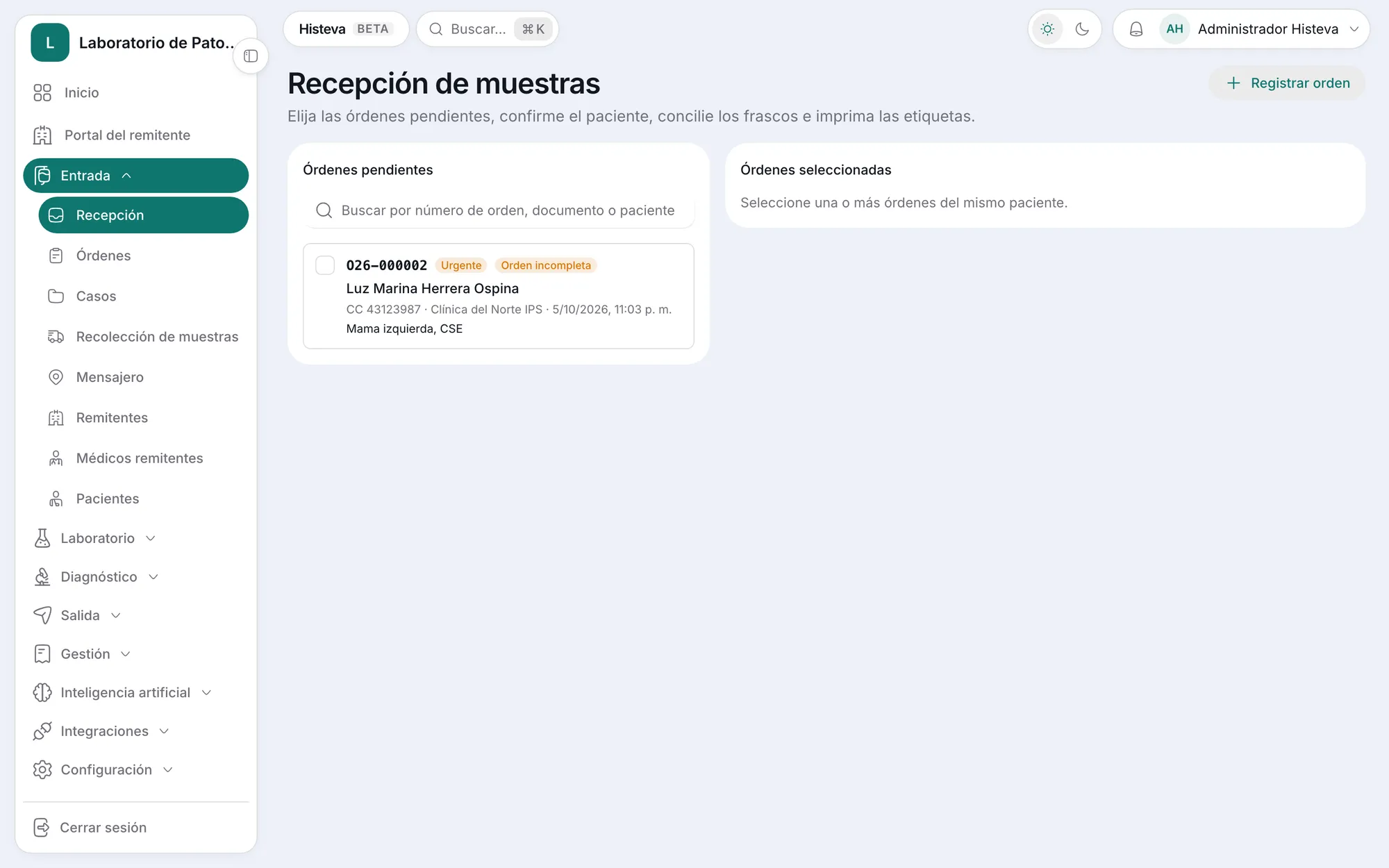
Since the code that links the image is the same one printed on the label, you don't need a scanner to get started. A lab can work at the microscope with Histeva from day one, with its slides labeled with DataMatrix codes, and add the scanner months later. Slides scanned from then on link to their cases automatically, with no data migration and no change of system.
This also changes the order of the investment: first you sort out identification and custody, which you need with or without a scanner, and then you buy the scanner with the workflow already in place. See pathology software.
What changes in the pathologist's day
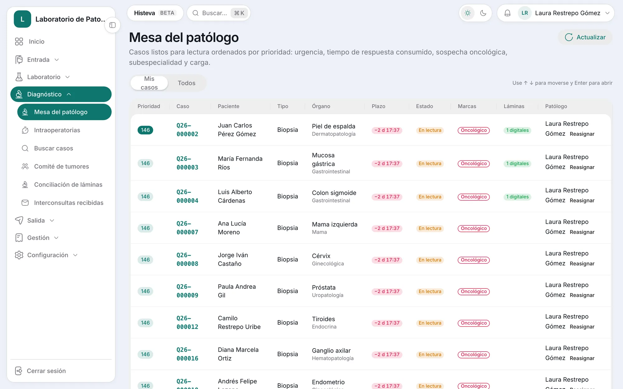
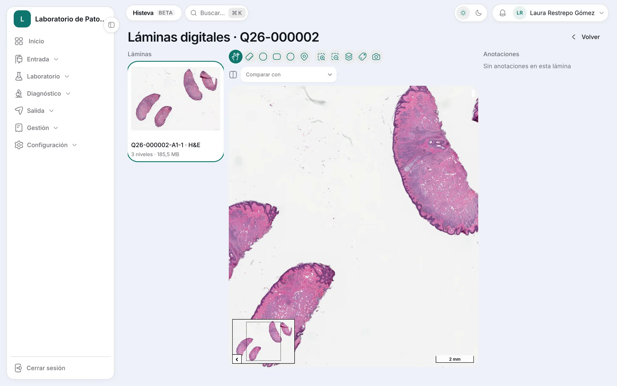
The pathologist worklist shows which cases have digital slides. When you open a case, the viewer lets you pan and zoom, measure, mark regions with annotations, and compare two slides side by side with synchronized navigation, for example the H&E against the immunohistochemistry. It also shows the overview image and the label image.
Consultations take a different form. Instead of sending slides by courier, you can share the case with a consulting pathologist through limited access to that case's studies, and their signed response comes back to the case.
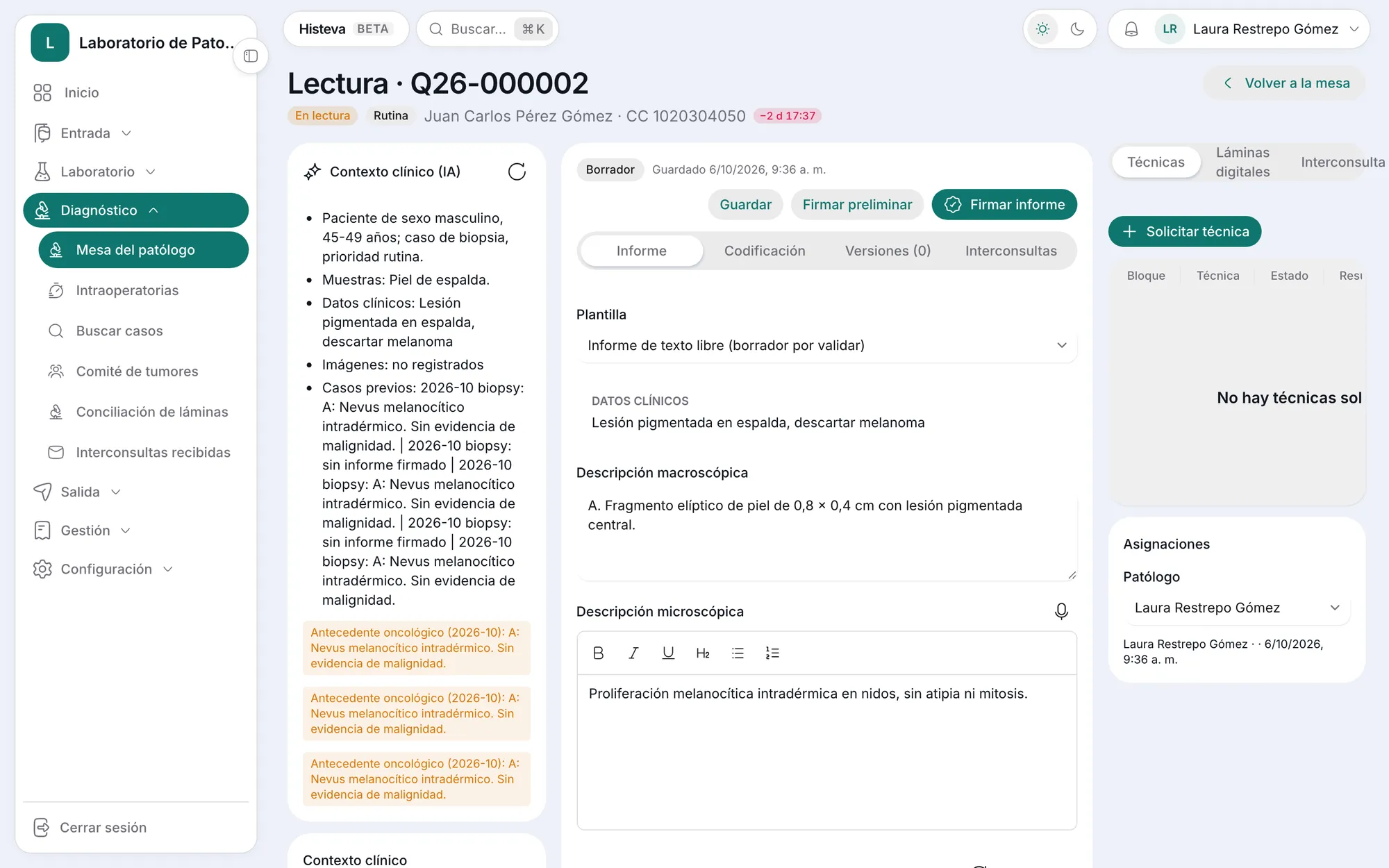
The report doesn't change: it's written, structured and signed out the same way, with or without digital slides. See synoptic pathology report.
On security: the viewer receives access permissions that expire after 5 minutes and renew while you work, and every image access is audited.
What it doesn't do yet
Image-analysis AI comes in a later phase. It will start with connectors to third-party models and, further on, a proprietary model once there are digital slides from partner labs. Today Histeva's AI works on text: dictation, synoptic drafts, consistency checking and coding. There's also no measured file size and upload figure yet for slides at 40x.
If you want to see how scanning fits into the rest of the workflow, take a look at how it works, pathology lab billing or book a demo.